Dynamic contrast-enhanced MRI of synovitis in knee osteoarthritis: repeatability, discrimination and sensitivity to change in a prospective experimental study
InflammationDCE

Inflammation
Bioxydyn has delivered multiple studies for pharmaceutical companies using quantitative imaging of inflammation in a wide range of diseases, including functional imaging of joints in rheumatoid arthritis and osteoarthritis and renal imaging in nephritis.
We use dynamic contrast-enhanced MRI (DCE-MRI) to quantify changes in perfusion and endothelial integrity in the presence of inflammation and during treatment. Bioxydyn has defined many of the standard approaches to the technique’s implementation in clinical trials.
DCE-MRI involves the use of standard clinical intravenously-administered gadolinium-based contrast agents. The uptake and washout of the agent is interpreted using physiological modelling methods to provide imaging biomarkers of microvascular function and inflammation.
Bioxydyn uses the Ktrans quantitative biomarker, which can be readily compared between centres. Its relationship to the intensity of inflammation is based on sound and widely accepted science, is extremely sensitive to drug effects, and only requires small patient numbers.
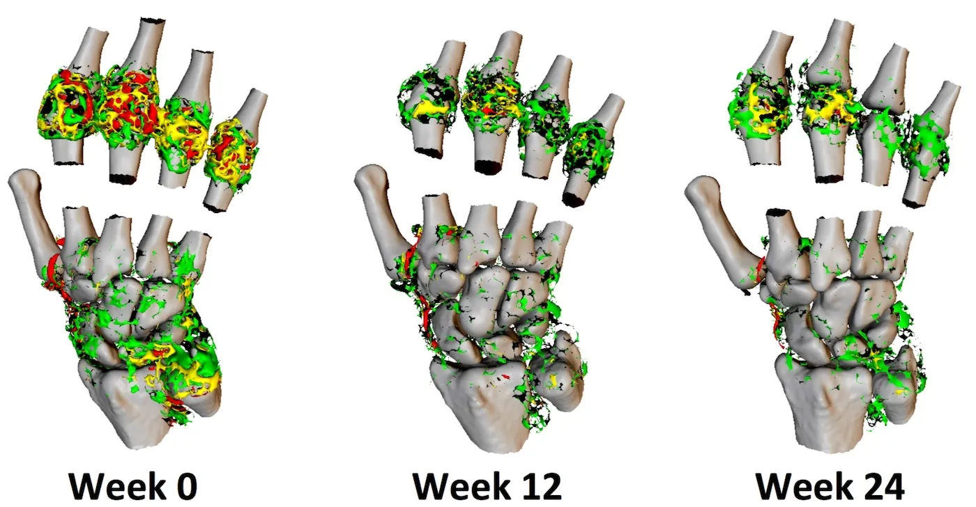
The images show change in Ktrans in the joints of the hands and wrist in an individual with rheumatoid arthritis during therapeutic intervention.
Renal imaging
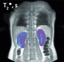
Bioxydyn is also experienced in using MRI biomarkers of inflammation and fibrosis in kidney disease. Examples include measurements of perfusion derived from arterial spin labelling (ASL), relaxation time measurements (T1, T2, T2*), volume measurements, diffusion MRI (ADC, FA). We are also experts in methods to measure hypoxic status (BOLD, oxygen-enhanced MRI) and pH (CEST).
The image shows maps of T2* in the kidneys of an individual with lupus nephritis. From Skeoch et al 2017.